
Following on from my post about the problematic field of mitochondrial transplantation, another paper was bought to my attention, in which the authors injected mitochondria into the eyes of human patients. The paper is posted as a pre-print on Research Square, which is Nature Publishing Group’s pre-print platform for papers under review or in process at their journals.
Leonid Schneider has a nice write-up about how this study was apparently funded – by the billionaire Bill Ackman (yes the republican mega donor, the same one who bankrolled David Sabatini). Just for good measure, Leo also covers another mito’ transplant paper which was missing from my previous analysis, with Sholto David finding more than a few numerical errors in the data. Add it to the pile of accumulating garbage that defines this whole field.
Dodgy billionaire funded pseudo-science aside, many of the problems I already outlined about mito’ transplant are present in this new paper, including an apparent claim that mito’s were counted using a hemocytometer (not possible). The mitochondria were isolated using the highly problematic serial-filtration method – the one that makes a kind of mito-ish soup with lots of blood contamination plus some residual BSA and bacterial protease for good measure.
Notably, the results section (page 7) of the paper claims that the yield of mitochondria was as follows…
“Hemocytometer counts gave 7.5 x 10^7 mitochondria for the first product, administered to the right eye, and 2.5 x 10^7 for the second, administered to the left”
So, the average total yield was ~5 x 10^7 mito’s (average of 7.5 and 2.5). Suspending all of these mito’s in 100 microliters (0.1ml) of buffer would result in a working concentration of 5 x 10^8 per ml. Later on in the methods section (page 18), we learn that…
“The final pellet was resuspended in approximately 100 microliters of cold Solution A to a working concentration of approximately 2 x 10^9 mitochondria per mL”
How did the effective concentration of mitochondria magically go up by 4 fold (i.e., 2×10^9 / 5×10^8)? Could it possibly be that hemocytometers cannot be relied on to count mitochondria, and in reality the investigators don’t really know how many mito’s they’re injecting?
Other problems surround the volumes used for testing vs. administration to the patient. It appears only 50 microliters were injected (using a 1ml syringe, which a silly way to do this and carries a risk of air bubbles), leaving only 50 microliters for all the various sterility and mito’ function tests that were needed. That’s odd because…
“Rapid microbial detection was by STAT Gram stain on an approximately 100 microliter retention aliquot”
How did they suspend the mito’s in 100uL, inject 50uL to the patient, AND have 100uL left over for microbial testing, plus endotoxin testing, MitoTracker Orange staining, and compendial sterility testing?
Given the known impact of antibiotics on mito’ function (including fluoroquinolines), it’s also odd that they chose to treat the patient with such an agent (moxifloxacin) beforehand. The eye surface was also treated with iodine (kills everything), plus neomycin/polymyxin/dexamethasone ointment. Neomycin is toxic to mitochondria. So is polymyxin. Vancomycin was also applied (you guessed it), along with ceftazidime and voriconazole. It’s a wonder there were any mito’s left after encountering this soup of antimicrobials.
Track Record
The first author of the new paper was also lead author on a previous clinical trial for mitochondrial transplant in acute cerebral ischemia. Naturally, that study prepared mitochondria using a “validated protocol” – the serial filtration method in the 2014 JoVE paper.
Notably in the ischemia study they measured mitochondrial viability using a Resazurin Cell Viability Assay Kit (from Biotium). This is nothing more than a simple NAD(P)H oxidase kit, essentially the same as an MTT assay. Apparently the kit was used “following the manufacturer’s instructions”, which is odd because the manufacturer doesn’t say anything about using it for isolated mitochondria instead of cells.
Of course, the paper shows a nice picture of some mito’s under electron microscopy…
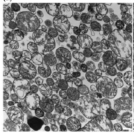
…and then proceeds to claim “minimal contamination from non-mitochondrial components”, on the basis of absolutely no quantitative measure whatsoever.
The other mito’ data are similarly crap. There’s an N=1 for TMRM staining. As already discussed they measured ATP in the prep, finding a concentration of 3 pmols per microgram. This apparently “aligns with expected viability benchmarks for isolated mitochondria” with a reference to this paper. The values reported therein are 11 for brain mito’s and 0.15 for liver mito’s, but there are no data for muscle mitochondria.
Somehow, “our muscle mito’s had some random amount of ATP that’s somewhere in the middle of what people found 25 years ago in mito’s from completely different tissues” doesn’t have the same ring of authority to it.
I think the only sane response to this latest example of mito’ transplant insanity is that I would rather stick pencils in my eyes!