The Guy Foundation & Quantum Biology – No Mitochondria do not Communicate With Light!

Quote

I’ve been meaning to write about this for a while, but what with running the lab, teaching, trying to stay on top of the poly-crisis of the NIH funding situation, starting an MBA, and doing my best to avoid GenAI wherever possible, it got put down the priority list. That’s unfortunate because this is a fun story…

If you work in mitochondria circles, you may have heard of the Guy Foundation?  It’s a charitable trust in the UK – a philanthropic effort from Geoffrey Guy, who made his money as co-founder of GW pharmaceuticals, the first company to get cannabis derived medications approved for human use. GW was sold to a US company for $7.2bn in 2022. Dr. Guy seems like a decent… guy… (sorry), and let me be clear this post is not intended as a personal attack on him, the company he founded, or the mission or operations of the Guy Foundation (GF).  Rather, I want to highlight the absolute bonkers batshit crazy science the foundation is spending its money on.

Flashy Science

Let’s get right into it, with the idea that mitochondria can communicate using light, explained in a YT video from Dr. Guy himself. Building on the widely accepted idea that mitochondria run on electricity (something, something, membrane potential, millivolts – go read Peter Mitchell’s “little grey book”), the GF seems to think mitochondria produce photons as well.  In-fact, much of the “science” funded by the GF is on the topic of biophotonics – the release and sensing of photons by biological systems.

Aside: There are myriad examples of biological systems that produce light – firefly luciferase being most familiar. These have been extensively characterized at the molecular level and reconstituted outside the original organisms – all useful and well-documented science.  That’s not what this is about.

The paper that warrants the batshit label is this one. Published in that bastion of scientific excellence Frontiers in Physiology, it comes from the lab of Jimmy Bell at the University of Westminster in London. Bell and first author Rhys Mould are part of the Research Center for Optimal Health at Westminster. The work was funded by the GF, as proudly claimed on their website and noted in the paper.

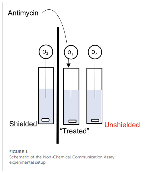
So what’s the problem?  Well, let’s start with a description of the experimental set-up from figure 1 of the paper: 3 cuvets with stir-bars at the bottom, each containing a suspension of isolated mitochondria. The left cuvet is separated from the others by a piece of cardboard (the thick black line), while the one on the right is next to the central one (i.e., “unshielded”).  Into each cuvet is placed a needle-type O2 electrode, to measure O2 consumption by the mitochondria.  Let’s number these cuvets 1, 2, 3 from left to right.

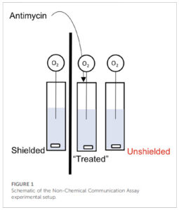
Now here’s the kicker… they add the mitochondrial inhibitor antimycin to the central cuvet (#2), and then claim the mitochondria in cuvet #3 respond by changing their O2 consumption (respiration), but the mito’s in the cuvet #1 do not. Not only that, the mechanism of communication involves mitochondria in cuvet #2 emitting photons and those in cuvet #2 sensing them.

Let’s ignore for just a second a couple of other minor problems with the methods, such as the use of 244uM antimycin (enough to tranq’ a horse) and the complete lack of any added metabolic substrate to support the mitochondrial respiration.  Let’s instead focus on section 3.4 of the discussion…

“3.4 Comparing “light” and “dark” experiments. Finally, we compared the change in OCR between unshielded mitochondria in light conditions versus dark conditions, shown in Figure 6. In MCF7, the rate of OCR change was significantly higher in light conditions compared to dark conditions.”

OCR here stands for “oxygen consumption rate”.  In other words, this magical communication between cuvets via photons was actually more pronounced when the system was flooded with gazillions of photons from the room lights, versus in the dark – exactly opposite to how low-intensity light phenomena are supposed to work. Batshit is not nearly a strong enough descriptor.

Yeah yeah nit-picky details, what’s the real problem?  Dear reader, take a look again at Figure 1 above.  Do you see any type of lid on the apparatus, or anything separating the liquid part (blue) from the air above it?

The problem is, the way to measure mitochondrial respiration / OCR / O2 consumption, is in a CLOSED system such as a Clark type oxygen electrode, of which there are numerous vendors available, or alternative platforms. Every one of these instrument makers will tell you that the liquid in which [O2] is measured (e.g. a suspension of mitochondria or cells at the bottom of a well) MUST be sealed off from the atmosphere… even the tiniest air bubble will mess up the readings.

This is simple chemistry – air is 21% oxygen – much higher than the solubility of oxygen in water (about 200 micromolar at 37C). If there’s any contact between the liquid and the air, any O2 consumed by mitochondria in solution will be instantly replaced by that from the air, so the concentration of O2 in the liquid will stay the same. It’s the CHANGE in O2 concentration that’s used to determine the RATE of O2 consumption (that’s the “R” in OCR).  No change in [O2]?  No rate.

Simply put… it is damn near impossible to measure OCR in an open system such as this (well, technically it’s possible using precise mixing at the gas-liquid interface, a complex series of engineering formulas, lots of math, and a full understanding of the problem just described, but that’s not what happened here).

Show Me The Data

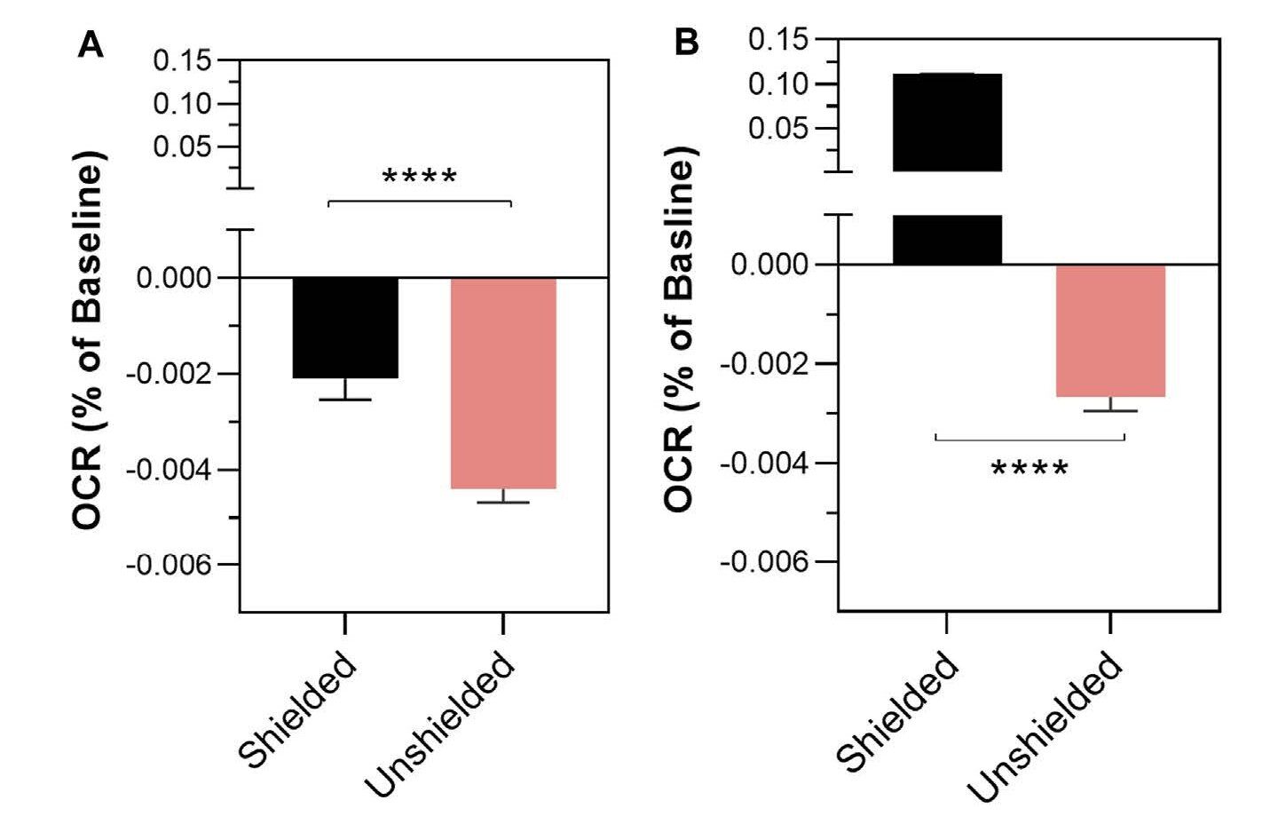
The “data” in the paper are presented in a manner completely opaque and unfamiliar to anyone who does these types of experiments.  The change in oxygen consumption rate (remember – there is no rate) in cuvets #1 and #3 following antimycin addition to cuvet #2 is shown in Figure 2…

Your eyes do not deceive you.. there was a 0.004% change in OCR in the cuvet that was exposed to a big old dose of pew-pew mito lazer beemz (#3, pink bar) but only a 0.002% change in the mito’s that were shielded by a bit of cardboard (cuvet #1, black). Tip – remember the direction these bars are pointing, for later on.

Far be it from me (a non-statistician) to wonder at the levels of intricacy and convolution required to detect a 0.004% change in anything, but this does not seem to be: (A) at all measurable, (B) of any importance. For context, this is like someone who earns $100k a year getting a $4 pay cut.  It should therefore come as no surprise that the statistics section of the paper will also appear odd to anyone familiar with such experiments…

“Differences in the rate of mitochondrial oxygen consumption
were analysed using a mixed linear effects model written in R”

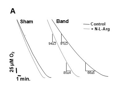
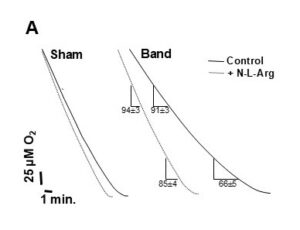
Complete overkill. This is not how it’s done. You calculate the rate (slope of the O2/time trace) and (after checking for normalcy) use simple statistical tests to see if the values are different, with appropriate multiple-testing corrections if necessary. If you want to get fancy you could even use the real-statistics plug-in for Excel, but you absolutely do not need a mixed linear effects model to see the difference between 2 diagonal lines on a trace. Here’s an example from one of my very old (fuzzy) papers.  This is what real oxygen consumption traces look like.  You can see that one line is different from the other.  Compare this to what you’re about to see further down the page.

Show Me The Data – Part 2

As a polite science critic, my first course of action upon reading this paper (well, OK, not my first choice) was to make a nice post over on PubPeer, bringing up the above points and more.

To the authors’ credit, they did respond to clarify some of the experimental details, but in some cases this only confirmed my concerns (e.g. there was a lid made of parafilm, but still a head-space, and indeed the buffer contained no fuel for the mitochondria to respire on). Most importantly, the authors were gracious enough to share the original data set on FigShare.  Absolute stellar move. Kudos. Love it. I’ve been using FigShare since 2016 and everyone should do this!  The problem is when you actually plot the data from the Excel sheet, as predicted from the impossibilities discussed above, there is no rate.  The concentration of oxygen is flat over time, because it’s at equilibrium with the atmosphere.

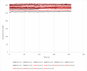
The y-axis here is dissolved oxygen (in micromolar, you’ll note it’s a bit higher than the usual 200 uM, likely because these experiments were done at room temp’ and O2 solubility is an inverse function of temperature). The x-axis is time in seconds, and the antimycin A was added at 120s. The black lines are 8 samples from the unshielded cuvet, and the red lines are the shielded cuvet.

Put simply THERE IS NO DOWNWARD SLOPE, THERE IS NO OXYGEN CONSUMPTION RATE TO SPEAK OF. Some of the lines even slope upwards, so the [O2] in solution is actually increasing over time.  If we calculate the rates before and after the antimycin addition, the numbers (mean +/- standard error) come out as follows:

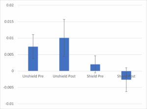
The chart shows the average rate (N=8 per group) for the unshielded cuvets (left 2 bars) vs. the shielded cuvets (right 2 bars), pre vs. post antimycin addition to the central cuvet. There is no difference between bars 1 and 2 – if anything the slope of the line (which is already close to zero!) increases instead of decreasing as claimed in the paper (compare this to Figure 2, above). The error bars (standard error of the mean) also show that whatever difference there may be is statistically non-significant – no fancy linear mixed regression what-have-you.  Looking at the unshielded vs. shielded groups at baseline (bar #1 vs. #3) you could even say they show a bigger difference than pre/post antimycin.  In other words, regardless of any mito’ communication voodoo, having a bit of cardboard next to you causes your O2 consumption to drop from very close to zero, to very actually zero, before any mito’ poisons are added to your neighbor.

Furthermore, the control data (cuvet #2) are not included, so we don’t even know if the massive dose of antimycin did anything.  It probably inhibited the mitochondria, but there would be no change in [O2] because, as explained above, any O2 consumed by the mito’s would be replaced by that from the atmosphere, so the trace would remain flat.  The difference between a flat line and a flat line, is nothing.

Anyway, I’d encourage anyone who enjoys playing around in Excel or R to take a look at these data and produce a single shred of evidence that the main conclusion is valid.

Let me be clear – not only is the key claim of this paper utterly bonkers, it’s based on a total lack of evidence, with a complete misunderstanding of how to measure mitochondrial function.

Surely this should be retracted?

Of course it should!  But since when did doing the right thing matter for publisher Frontiers? (just ask Leo Schneider). I wrote to the person who edited the paper, YoungChan Kim at the University of Surrey, UK, but he didn’t bother to respond. Neither did John Imig the chief editor for this field section of Frontiers in Physiology. A good friend who is involved with the journal did respond to my inquiry, but they were not involved with handling the paper and so could not do anything about it.  There was a bit of chatter in the PubPeer thread, which died down as soon as I posted a “no rate” graph similar to the one above.

Importantly, David Fernig raised the point that this is not an impossible experiment to do properly. You just need a very sensitive photomultiplier tube to detect photons, and tightly controlled other conditions (sealed chambers, dark room, temperature regulation, filters to control the wavelengths of light passing between samples, etc).  Maybe this IS something worth investigating?  Maybe the GF should give the money instead to someone who understands this?

How will this “knowledge” be used?

The paper is still out there, and people are still using it to make ridiculous claims.  The GF is lauding the work in their newsletter, as well as the aforementioned video, the lead author also talks about it in video format.  The foundation appears very proud of the work it funded, which is unfortunate because it’s not very good at all.

Of course, if you believe mitochondria can influence each other via light, it’s a small step away to hack that biology by shining bright lights on various body parts, which brings us neatly into crazy anti-aging therapies, such as the claims that red light can energise your mito’s. I’ve made my thoughts on the shit-show that is anti-aging biotech abundantly clear before. While there are certainly biological effects that can be attributed to certain wavelengths of light, they are usually based on rigorous experimentation (such as the paper just cited, from a well-known group in the field at MCW). The Frontiers paper is not an example of quality science.


There’s a lot more to be written on the various out there projects and papers claiming all sorts of weird quantum effects on mitochondria. It’s not just light… there are all kinds of papers about electromagnetic fields and mitochondria.  Claims that mitochondria operate at high temperatures inside cells. Mitochondria allegedly respond to music and other sounds.

But for now, we just have to wonder how does a charitable foundation hand over non-trivial amounts money to fund “pew-pew mito space photon lazer beemz”, and then when the project execution and results are complete garbage, they just accept everything at face value?  No admission that anything silly happened.  No acknowledgement that the work is absolutely fundamentally flawed, and was not performed in a way that could give meaningful outcomes. It’s an alternate reality!

Spring 2025 Update

It’s been a while! Several updates to the lab over the past few months…

  • Rahiim Lake passed his PhD qualifier exam last fall (congrats!)
  • We hosted the 11th annual TRiMAD (Translational Research in Mitochondria Aging & Disease) conference here in October, attended by over 160 people.
  • Paul spoke at a number of conferences and outside institutions, including a conference at UPenn on research integrity, resulting in this paper.
  • The University finally adopted and published the Open Access Publishing Guidelines that we worked to bring through the faculty senate. It only took 7 years!
  • Paul went back to school in the Executive MBA program at UofR’s Simon School of Business (anticipated graduation May 2026). It resulted in finally (reluctantly) getting a linked-in page.
  • Post-doc Sabarna Chowdhury got married in April 2025 (congrats!)
  • Lots of other papers got published, including some exciting work on taurine metabolism with the Bajaj lab in the (newly NCI designated) Cancer Center, which is accepted for publication in Nature.

Buchi Rotavap Parts

A rotary evaporator (RotaVap) is a core piece of equipment for any lab that does chemical synthesis, and Buchi make some of the best ones. Unfortunately, ours is about 25 years old and they no longer make parts for it.

In the heart of the apparatus there’s a worm-drive… the motor is connected to a helical spindle which drives a ring gear. This is what spins the round-bottomed flask.

Over the past few months we’d been noticing that at low spin speeds, the rotation would skip.  It would intermittently speed up and slow down through the rotation cycle.  So, we opened up the gearbox to see what was inside (note – you may need to use a hammer to get the parts to separate out).

The problem is, Buchi decided to make the gear out of plastic, and years of exposure to grease and chemicals causes the plastic to crack, as shown here…

The result is the motor spins but the plastic rung just grinds around and doesn’t spin the metal part that’s eventually attached to the flask.  The worm gear is basically slipping once per rotation. We tried buying a used model off eBay, and upon dissection it had exactly the same problem, so it appears this is a common fault on this particular model (RE-111).

The solution is not as simple as “glue it back together”. There’s too much grease everywhere, so it needs completely stripping down to remove all that so the glue will stick. And then, removing the ring gear can only be done by snapping it in two. The problem then is there’s not much surface area to glue back together so it will probably break again soon.

Enter the 3D printer!

With some quick work in Sketchup  I was able to make a model of the gear with a break in the middle and plenty of surface area for gluing the 2 halves together, as well as gluing to the metal spindle inside the machine.

Here’s the new gear, printed in Elegoo ABS-like resin on a Mars 3 printer. The STL file is here to download if your Buchi Rotavap is in need of a fix!


Spring ’24 Update

Lots of happenings in the lab since last fall:

  • Grad’ student Rahiim Lake (Biochem Grad Program) joined in summer 2023 and is getting up to speed on his project looking at MGO modification targets in mitochondria.
  • New Post-Doc’ Sabarna Chowdhury arrived from India in January and is learning the various methods of the lab.  Sabarna is a zebrafish biologist by training, but also a bona fide mitochondriac!  She will (hopefully) be attending the CSHL course on metabolomics this summer.
  • Our paper on ROS generation by reverse electron transfer at complex I was published in Redox Biology.
  • Paul is now back on the Faculty Senate’s Research Policy Committee (RPC), which this year will be reviewing several policies including the one on research misconduct. Yummy!
  • We continue to fight misinformation and bad science, especially in the field of mitochondrial biology.  Most recently this has included calling out a truly awful study claiming that mitochondria can communicate with each other across distances of centimeters by using photons!
  • A successful Biochemistry retreat was held, in which we hosted Rita Alevriadou from University of Buffalo, who gave a fascinating talk on mitochondria inside extracellular vesicles.

Resurrecting a Seahorse XF96

Those familiar with Seahorse XF instruments are aware that since the company was bought out by Agilent, there has been a tendency toward “sunsetting” older instruments and encouraging users to upgrade to newer models. Most recently, this came in the form of an email claiming that, not only would the older XF96 instrument be no longer supported, but also the availability of consumables would cease…

Naturally, Agilent’s preferred solution to this problem is for the end-user to drop $100k+ on a newer XFe96 or XFpro machine. For those like myself without deep pockets, this is not an option. We’ve used Seahorse products for a long time (in-fact we had one of the very first XF24 devices sold), but we’re not in a position to drop that kind of money every 5-6 years just to keep doing our experiments.


Crappy PCs

Adding to the problem of the instrument itself becoming obsolete, the XF96 shipped with a “POS” terminal – a piece of shit touchscreen 32-bit PC running Windows 7 with a measly 2GB of RAM. The touchscreen interface never lined up properly, and simply added to the overall slowness of the machine (here’s the underside of the offending PC – yes those are RS232 serial ports).

In addition to frequent crashes (sometimes during the middle of experiments!) many universities including my own will not allow older PCs such as this to connect to their network, so this effectively renders the PC incapable of receiving security upgrades.The use of USB thumb-drives to transfer data on/off the PC is therefore risky.


Simple – Just Swap in a New PC!

You’d be mistaken for thinking the solution here is to simply swap in a brand-new PC and transfer the software. That’s because there are a number of software and hardware dependencies which anchor the XF to the VERY specific PC it ships with…

  • The XF communicates with the PC via two separate cables: an old-school RS232 serial port for the instrument itself, and a separate USB/serial port adapter for the barcode reader.
  • Multiple subroutines within the XF software need old-school plug-ins and dependencies. This includes Adobe Flashplayer, MS Edge plugins, Visual C++, and running most things in compatibility mode for Win7.
  • The system needs a VERY specific version of Microsoft Excel 2010.

In-fact, in addition to some minor tweaks regarding temperature control inside the instrument, one of the key advantages for the XFe96 versus the XF96 as marketed by the company, was the ability to run on a modern 64-bit Win10 PC. In effect, they crippled the XF96 so badly you couldn’t even upgrade the PC connected to it!


Sounds Like a Challenge

Naturally, not wanting to continue running our XF96 on a defunct old PC, I undertook the challenge to get a modern Win11 PC speaking to it. The PC I chose was this cheap fanless mini-PC that sticks to the back of a monitor. It comes with Win11 preinstalled and 6 USB ports, so plenty of room for expansion. Since it doesn’t have any serial ports, I also picked up another USB/serial adapter cable.

Below are the steps to install the relevant software. Note that many of the files are no longer available through official channels such as Microsoft, so you need to find a link (e.g., from an old discussion forum) and then copy the URL into the Wayback Machine, then visit a cached version of the old link. It goes without saying this is all at your own risk, with no implication here that any of the linked resources are reliable, virus-free, or whatever. There’s also some light registry editing involved, which comes with a FAFO disclaimer!

  • First install Microsoft Visual C++ redistributable 2010. Even though most new PCs are 64-bit, you will need both the 64-bit and 32-bit (x86) versions of this. I obtained these files here, but you can also find them using the cached link method as described above.
  • Download and install Java.
  • Download and install Microsoft Edge WebView Runtime X64. This should come via Windows Update on a modern PC, and it replaces the Active X module in Internet Explorer that was required on the original Seahorse PC.
  • Install the XF96 installer package. The file is no longer available from the Agilent website, but the one you’re looking for is “XF96SetUp.exe” and the Software version is 1.4.2.3 from 2018. DO NOT install the “Wave” software, as this will not play nice with the XF96. Hopefully any lab with an existing XF96 machine should have a copy of the software installer sitting around.
  • Install Microsoft Excel 2010. If missing the original DVD, you will need to find an ISO or EXE file to download. You will also need a Key Extractor to recover the software key for Excel from the old PC (see below). This site lists all the links for Office downloads, and the file you’re looking for X16-30329.exe from this link (need to use Wayback to find a cached version). **You can’t use any Office 2010 ISO file. It has to be from the retail version of Office Pro, NOT the pro-plus version which was for enterprise distributions. If you install the wrong version, the recovered software key will not work.
  • Download and install the driver for the original USB/serial cable that came with the XF96. This should be a Gigaware device, and will install the file “Svk2pl64.sys” in the folder C:\Windows\System32\Drivers\ You may also need another driver for the additional USB/serial adaptor purchased to go with the new PC.
  • Extracting the software key from the old copy of Excel is not something Microsoft wants, so key-extractor software tends to reside in dark corners of the internet.  Tread carefully – such software WILL trigger a virus warning and quarantine on a modern PC, so download and transfer it to the old PC as a .zip file and then only unpack it on the old PC. Remarkably, in my case doing this did not trigger a virus warning on the old PC – a good sign that it’s not suitably protected!


HARDWARE SETUP

You’d think the simple approach would be to use the old black Gigaware cable for the barcode reader on the old PC, to connect the barcode reader on the new PC, then use the fancy new USB cable to replace the serial/serial connection. NOPE – for some bizarre reason this doesn’t work (the barcode reader will connect, but won’t capture anything). The new cable must be used for the barcode reader.

  • Start with all cables disconnected. Plug the Gigaware cable into a USB port. Type “DEV” in the Start Menu and open “Device Manager”. Under “Ports”, double click on “Gigaware USB to Serial Cable”. Under the “Port Settings” tab, click “Advanced, and set the port to COM1.
  • Connect the other (serial) end of the cable to the XF port labeled COM1 (looking from the back of the machine this will be on the right).
  • Now turn on the XF96. Launch the XF Utility App (located in C:\Program Files (x86)\Seahorse Bioscience\XFReader96\Utility\). Under the “Connections” tab, select COM1, and click connect. Successful connection yields a green light. If prompted for a passcode, use “OSTER”.
  • With the utility app still running in the background, now connect the second (new) USB/serial cable to a different USB port. Open Device Manager again, and use the same procedure as above to assign this cable as COM2.
  • Then connect the other end of this cable to the port labeled COM2 on the XF (looking from the back of the machine this will be on the left).
  • Finally, in the Utility App, in the “Barcode” tab, select COM2 and connect to the barcode reader.


TRANSFERRING SETTINGS

Every Seahorse XF machine is slightly different, so a bunch of instrument-specific settings are stored in the registry for call-up by the software. This includes the offsets for temperature regulation, and the precise positions of the motors for different functions. All these settings have to be transcribed over to the new PC. There are 3 different ways of doing this…

(1) Before closing out the original PC, export a copy of the registry settings containing all the parameters from the old machine. These are stored in HKLM\SOFTWARE\WOW6432Node\Seahorse Bioscience\XF96_CTRL

(2) Alternatively, before disconnecting the old machine, launch the Utility App and take screenshots of all the tabs (especially Motors). Or just write down the motor positions and other important numbers from the Utility.

(3) A third approach is in the main Seahorse software itself – navigate to the “Instrument Setup” screen and click the “Administrator” button (right side). On the left, click “Hardware Settings” – a password prompt will appear (PW: GENIUS). Then click the various tabs to note down important parameters such as probe positions.

Once you have all the settings noted, keep them in a safe place. Then open the Utility App or the main Seahorse software on the new PC and enter all the values. Or just edit the registry. It is essential at this point to restart both the XF machine and the PC, and make sure all the settings have “stuck” in the registry.

*Oddly, when set-up was all finished, the Seahorse software would only recognise that there were 2 injection ports, not the usual 4.  There’s a registry setting “NumPorts” that was set to 2 – set it to 4 (decimal) and restart, then all the ports will be available.


IT’S ALIVE! (but what about consumables?)

If you’ve done everything correctly up to this point, you should now be able to turn on both the PC and the XF, and have them play nice with each other. At that point, it’s time for an experiment…

BUT… recall the original email from Agilent said that XF96 comsumables would no longer be available? This is not an immediate concern, because XF consumables never really go off (we’ve used cartridges 5 years past their nominal expiration dates with no problems). But, for those in need of new plates and cartridges, it appears the claim they are not available is simply untrue at this time (October 2023)…

There are two types of consumables on the Agilent website. This type (catalog #103798-100) is compatible with both XFe96 and XFPro machines, and we have found these cartridges and plates work perfectly well inside an old XF96 machine! There is also a newer line of plates (103777-100) that is ONLY compatible with the newer XFPro machine, and these won’t even work with the XFe96. Don’t buy those ones.

It’s notable that very recently the XFe has also disappeared from the Agilent website, so one imagines they are planning to phase that out soon as well. But, in the mean-time, it appears that plates/cartridges marked as XFe96 compatible will work on the XF96.

It should also be noted there are additional new plate types (e.g., spheroid/organoid plates), wherein the plate itself is a completely different shape, and these physically will not fit inside the old machine. Apparently you can CNC-machine the metal block plate holder inside the machine so these new plates will fit, but I wouldn’t know anything about such shenanigans!

SUMMARY

It’s very possible to keep a ~10 year old Seahorse XF96 functioning, and have it talk to a modern Win11 64-bit PC, with all the advantages that conveys (virus protection, software and OS updates, networking, watching YouTube while your experiment runs).

The relentless march of planned obsolescence in scientific apparatus shows no signs of slowing, so I have no idea how long this particular set of hacks will buy us. Maybe a year, maybe more? Either way, for those without $100k+ to spare this is really the only option.

P.S. Used XF96’s can be had on eBay for $2-3k, so in theory with a cheap mini-PC you could build a working Seahorse system for well under 5 grand!