
Finally, our metabolomics paper is in press at J. Mol. Cell. Cardiol. (email if you want a reprint). TL/DR… SIRT1 drives most (~85%) of the metabolic alterations that occur in the heart during acute ischemic preconditioning (IPC).
This was quite a tough paper to get published. We started the project in spring 2013, and wrote it up in fall 2014. It got rejected from a big journal (IF>15) first, then went 2 rounds at a mid-level (IF>10) journal before being rejected again, and then it went 2 rounds at JMCC before acceptance. All told, a year of back and forth with reviewers and editors.
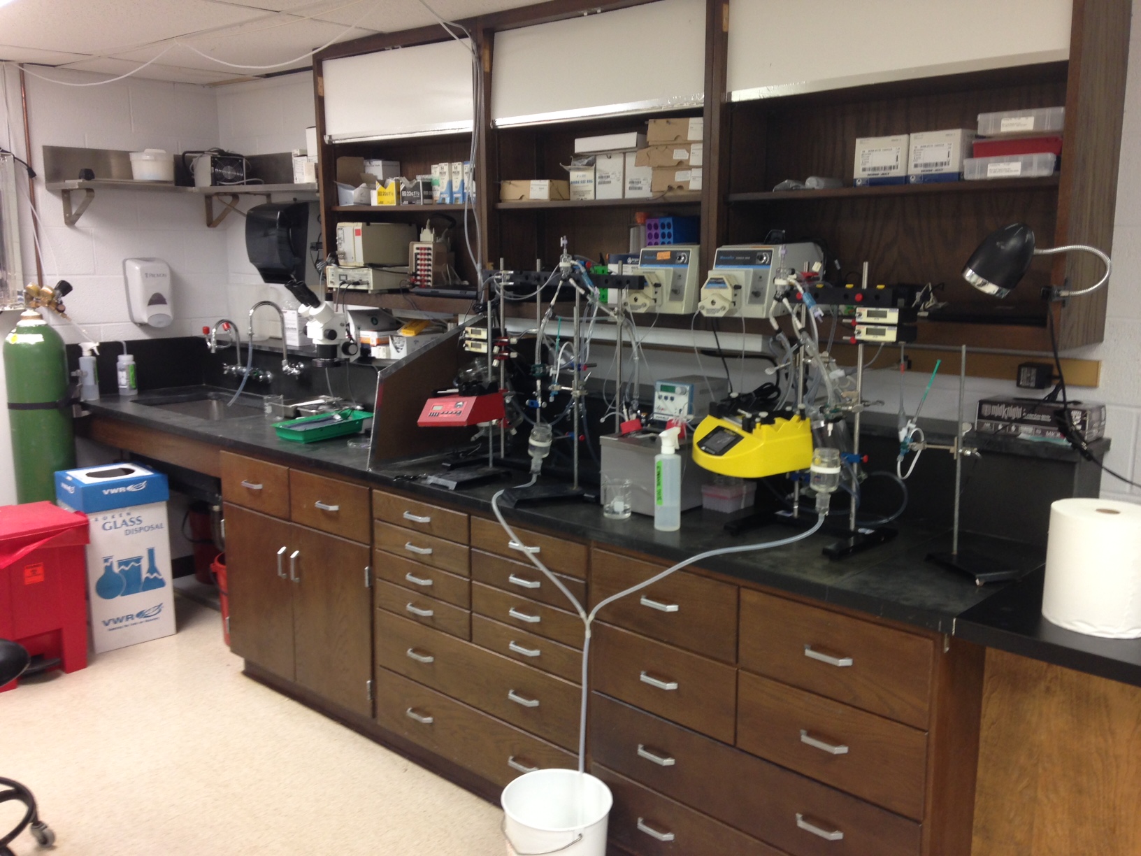
The model system we used to investigate this topic was the Langendorff perfused mouse heart and splitomicin, a pharmacologic inhibitor of SIRT1. The basic issue with the reviews that ended up as rejections, was an insistence by reviewers that we do things in-vivo and using knockout mice.
Normally, we’re big fans of moving toward more physiologically-relevant model systems, but in this case there are very specific reasons to use a perfused heart and a pharmacologic inhibitor. Here are some key points…
(1) Regarding pharmacology, the inhibitor we used is one we’d already shown can block acute IPC, so it’s a good candidate to test whether it also blocks the metabolic effects of IPC. Also, we had already shown that a SIRT1 KO mouse heart cannot be preconditioned, and that the endogenous protection seen in the SIRT1 over-expressing transgenic mouse can be blocked by 5 min. infusion of the inhibitor. Thus, the time-frame for the effects of SIRT1 in IPC is very short – on the order of 20 min. The SIRT1 KO mouse has known long-term metabolic alterations which would mask any changes we’d look for in IPC.
(2) Regarding in-vivo vs. in-vitro, it all boils down to sampling time. In our system, we can clamp the heart in liquid nitrogen Wollenberger tongs, straight off the perfusion rig. In effect, it goes from beating to frozen in less than a second. That’s important for getting reliable information on metabolites such as ATP, NADH, GSH and other labile redox things.
The problem is, when you precondition a mouse heart in-vivo, it’s a focal ischemia model. Only part of the heart is ischemic (the bit downstream of the vessel you occlude), so if you try to dissect out the ischemic zone, you delay the clamping by a couple of minutes and destroy all the labile metabolites during the dissection. Alternatively, if you clamp the whole heart right out of the animal into liquid nitrogen you create 2 problems… First, all the changes in the ischemic area get “diluted” with the other part of the heart that wasn’t ischemic (the so-called “area not at risk”). Second, you’re also sampling blood, so you don’t know if the changes you see are in the myocardial tissue or the blood that comes along for the ride (by our estimates when you clamp a heart out of a mouse, about 1/3 of the sample is blood). In contrast, the perfused heart system has no blood, so the whole sample is myocardium. Also the entire heart is ischemic, so there’s no dilution.
(3) The other major issue concerns the type of metabolomics analysis you want to perform. In this paper, we performed not only steady-state metabolomics (i.e., measuring the relative levels of metabolites), but also 13C labeled substrate tracing. The latter can yield proxy information about metabolic flux, which steady-state measurements cannot. This is easy in the perfused system… just throw 13C-glucose or 13C-palmitate in the perfusion media, but in-vivo this creates problems. You can’t just deliver labeled substrate to a whole mouse and assume it’s only being metabolized by the heart on first pass. For example, the cardiac/liver Randle cycle can result in labeled glucose being turned to labeled fat by the liver, then sent to the heart as fuel. Also, whatever 13C-substrate you infuse is going to compete with endogenous blood-borne substrates in the animal. In the perfused system you can swap out the whole substrate (i.e., replace all the glucose with 13C-glucose), so you have much tighter control over delivery.
So, this really is one of those cases where an abstract application of Krogh’s principle comes into play. The in-vitro and pharmacology based approach really was the best system available to answer the question at hand (that question being, what fraction of the metabolic changes that occur in acute IPC are governed by SIRT1 signaling?)
Naturally, we argued all the above points and it didn’t get us anywhere! As a lab that routinely uses both in-vivo and knockout models, it’s rather frustrating to be locked out of publishing in certain journals because we chose to use an allegedly inferior system. It’s annoying that some journals have a myopic focus on knockouts and in-vivo data which precludes them from publishing otherwise solid work. Thankfully JMCC seems to have a more sensible approach to this type of work!