Fall 2021 Updates

COVID

A year and a half into the pandemic, and with the benefit of 20/20 hindsight, I will gladly admit that we fucked up badly by culling our mouse colonies back to a fifth of their normal size in March of 2020 – it’s taken us over a year to get everything back up to size and running again, and meanwhile everyone else just #DGAF and carried along doing mouse work. Ugh!

Still, I get to take out my rage by shouting obscenities at the moronic antivax protesters outside the hospital on my daily bike ride. I should really invest in a Super Soaker filled with MMTS to spray at them.

PAPERS

(1) Our paper with Ana Jimenez of Colgate University, on metabolomics of young and old small and large breed dogs, was finally published in Geroscience. This was part of a larger study by Ana, aimed at addressing the question of why large dogs die prematurely (which is opposite to most of the rest of biology, where large animals live longer). The graphical summary below highlights some key differences in metabolite abundances between fibroblasts from the 4 different groups. Of particular interest (to me) is that carnitine seems to go up as small dogs age, but not large dogs, which might suggest some differences in ability to burn fat, which could be targeted pharmacologically in older dogs.

(3) Our paper on a modified blue-native (BN) gel method was also published, as part of the Methods in Molecular Biology book series. Blue-native is a method for looking at large protein complexes, including so-called “supercomplexes” (SCs) of the mitochondrial respiratory chain. There’s been a lot of debate about the functional importance of SCs, and this paper is the result of a project to streamline the method to reduce a potential artifact source. The project itself was done by an undergrad summer intern, Megan Ngai, who is going to be starting medical school at SUNY upstate next year.

The premise of the modified method is quite simple… most BN methods involve treating your samples somehow, then extracting with detergent, and this step necessarily involves pelleting the mitochondria by centrifugation, prior to extraction. The problem is, doing so might select for “good” or dense mitochondria, and many of the perturbations that have been shown to alter SC formation are also associated with alteration of mito’ density (e.g. respiratory state, PT pore opening). So, what we did was to develop a compromise mito’ buffer that is suitable for both mito’ incubations/treatments and for extraction. This eliminates the pelleting step, so the mito’s that get put on the gel are not “sampled” in any way – they’re representative of the whole population. The upshot is that we find many perturbations of mito’ function simply do not pan out with differences in SC formation or abundance. That’s not to say all the data so far on SC’s is artifactual, but it does suggest that acute alterations in mito’ function really don’t seem to be linked to SCs in a functional manner.

FUNDING

The main R01 that funds the lab (HL-071158) is sadly now in no-cost extension, as the competing renewal application was reviewed in February but missed the payline by 2%. A revision was submitted in July and will be reviewed in October, so we are currently awaiting with fingers crossed. If the news is bad again, it will be the end of the line for this grant which my lab has held since 2003.

Our other funding is in the form of an R56 1 year award (at half modular budget) to gain more data for Aim 2 of the parent R01 application (DK-126659). That award is also in NCE, since the high-fat-diet studies that underpin it were delayed due to the mouse problems outlined above.

So, we’re running on fumes for the time being, hoping for a funding hit and trying to remain productive and optimistic in the interim. This cyclical nature of funding is nothing new, but it bites really hard when a grant you’ve had since your lab began (and which has produced over 100 papers in 17 years) might not make it.

PROJECT HAPPENINGS

  • We have successfully established a breeding colony of Glo1-/- (glyoxalase-I knockout) mice. The founders came from Jim Galligan in Arizona, and we’re using them to explore methylglyoxal stress in the Alkbh7-/- mice, as reported here.
  • On a related note, we tried last year to get a “matters arising” published in Nature, regarding a paper on lactylation of lysine residues. A pre-print of our letter is here, in which we point out 3 things. First, the MW of the lactyl-lysine moiety is the same as that of MGO-induced carboxyethyllysine, so mass spec’ alone cannot distinguish these PTMs. Second, the anti-lactyl-lysine antibody developed by the authors (which they sell through the PI’s company) appears to recognize CEL (or another MGO-induced adduct) better than it recognizes lactyl-lysine. Thirdly, the concentrations of lactate used to induce lactylation (25mM) are inhibitory to the enzyme GLO-2, such that they will elevate S-lactoyl-glutathione levels, and this is probably the underlying mechanism driving lactylation (as has been shown by Galligan). Unfortunately, after a year of dicking around Nature decided to reject our letter (and the author’s response) and the authors were quite rude to us about the whole affair, accusing us of stealing their ideas, and telling us to stop confusing people. All highly unprofessional, but par-for-the-course in glam-publishing land. Ugh!
  • A paper on ERK5 signaling and cardiac mitochondrial hibernation with Jun-Ichi Abe should be out in Redox Biology soon.
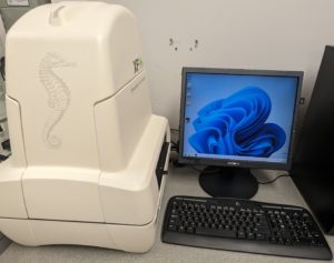
  • Grad student’ Alex Milliken’s project on measuring ROS in intact beating mouse hearts is progressing rapidly. We have built a Langendorff perfusion apparatus into a spectrofluorimeter, which allows measurement of reflectance-fluorescence of the heart, as shown in the picture below:
  • Using this system we can measure not just parameters like ROS, but also NADH, FADH2, membrane potential (TMRE) and mito’ PT pore opening. While others have done this in larger hearts (guinea pig or rat) this is the first such system in mouse heart, and also the only system with simultaneous measurement of cardiac function at the same time, with a pressure transducer balloon. Thus, we can relate changes in fluorescence to changes in cardiac function in real time.
  • We’re still working on ALKBH7, trying to figure out its biological function. In that regard, we were intrigued by a recent paper that claims to have discovered a role for the enzyme in regulating mitochondrial ribosomal and RNAs. To us, this is weird, because previously the crystal structure of the enzyme was specifically called out for lacking any binding sites for nucleic acids, as seen in some other ALKBH family members! The authors of the new paper seem to gloss over this by simply stating the enzyme has binding loops conserved from E. coli AlkB. But we’re not convinced the diagrams show any such conservation (rather, just a bunch of disordered structure). Adding to the complication, nothing about this new finding has any impact whatsoever on the underlying phenotype of the Alkbh7 knockout – namely, they don’t die of necrosis! Why would knocking out an RNA demethylase have any impact on acute necrotic signaling? So, this doesn’t really change much about our ongoing quest to determine how the enzyme is involved in necrosis, and how it interfaces with glyoxal metabolism (which the authors conveniently fail to mention, or cite our work). The proteomic data from the paper (which mainly uses siRNA knock down) also disagree with our own findins in knocout mouse hearts – namely they report large scale reductions in mitochondrial transcripts, but we saw no differences in any of the respiratory chain complexes, and no differences in mitochondrial enzyme activities. It could be this is highly cell-type specific, with ALKBH7 having an RNA-processing role in dividing/proliferating cells, versus more of a metabolism/cell-death role in terminally differentiated tissues such as the heart. Lots still to learn about this fascinating protein!

TRAVEL & CONFERENCES

We attended the AHA BVCS conference, where Alex presented a poster. We will also be at the Society for Heart Vascular Metabolism (SHVM) conference in later September, and the Society for Redox Biology & Medicine (SfRBM) conference in November. The latter is supposed to be in person, with Paul presenting at the sunrise free radical school, but I’d say with Covid on the rise, the likelihood of us going to Savannah GA any time soon is not looking high.

Hopes for in-person travel are slightly better however, for an upcoming Keystone Meeting on Mitochondria, Metabolism & Heart, in Breckenridge Colorado next February. Fingers crossed for less moronic behavior by the antivax population between now and the end of the year!

Publications Update

3 New papers to add to the lab publications list, to start the year on a good note…

  • This paper in Chem Med Chem with Paul Tripper’s lab (who are now based in Omaha Nebraska) reports on a new series of diazoxide derivatives, some of which are inhibitors of mitochondrial Complex II.
  • This paper in Autophagy with Keith Nehrke’s lab reports on the interactions between the hypoxic mitophagy receptor FUNDC1, and the mitochondrial unfolded protein response (mitoUPR) regulator ATFS1.
  • This paper accepted into JCI Insight (still in press so the link goes to the BioRxiv preprint) with URMC’s Michael O’Reilly and Dave Cohen, reports on the role of fatty acid synthesis in the development of the cardiomyocytes that extend along the pulmonary trunk (yes, the pulmonary vessels have muscle cells and they handle fat just like regular cardiomyocytes do!)

Going beyond faked data… faking the raw data!

Most readers here will be familiar with the issues surrounding western blotting as a technique in the bio-sciences, namely it is highly amenable to fabrication and inappropriate manipulation. A quick glance at PubPeer, ScienceIntegrityDigest, ForBetterScience, and other blogs reporting on scientific misconduct, makes it clear that blot-fakery is alive and well in 2020. Today, I will walk you through a paper I received for review which takes this to a whole new level!

What’s the problem?

One of the most common problems encountered with western blot data, is the presentation of “letter-boxed” blot images such as the one shown here…

In this image, 4 different proteins (ABCD) are blotted under 4 different experimental conditions (1234). These types of image are problematic for several reasons. First, there are no molecular weight (MW) markers, so it’s impossible to tell if the antibody recognized a protein at the correct MW. Second, only a small vertical slice of gel is shown, so it’s impossible to tell if the antibody only recognized one protein or several bands (how specific was the Ab). Third, in these types of collages the bottom blot (D in this case) is typically the “loading control”, showing the abundance of a house-keeping protein such as GAPDH or beta-actin, thus allegedly proving that equal protein was loaded in each lane. The problem is, as you can see from the example above, the spacing, size, shape, and slope of the bands is different in each blot. This makes it clear that the “loading control” likely did not originate from the same blot membrane – the authors simply loaded a different gel and blotted that, so this is not a true loading control. Fourth, western blotting as a method suffers from a woefully narrow dynamic range (about 10-fold), so it’s important for quantitation purposes that the bands be exposed somewhere within that range. Here, all of the bands have solid black centers and are overexposed, so the bands are completely useless for quantitative purposes. Lastly, the background (more on that in a minute) is plain and washed out, so it’s impossible to “anchor” the bands to any background features. This makes it impossible to tell if any of the bands have been pasted or spliced together. It doesn’t mean the image is fake, it just means it’s impossible to say it’s not fake.

When I receive a paper to review, if the bulk of data is presented in this letter-boxed format, I will often reject the manuscript, or demand better quality evidence.

How have publishers reacted to the blot-fakery crisis?

In recent years, many journals have become aware of the shit show that is blot-fakery, and have started to demand better quality controls for submitted manuscripts. This typically requires provision of full-sized, uncropped blots as original images. Since most journals are online and storage is not an obstacle, this is now easy for authors (of authentic work) to comply with. Many publishers are going a step further and demanding complete original data sets behind every figure! My lab has been doing this for a while now, posting complete data sets on the file server FigShare.

How have faking authors responded?

One might think that simply demanding better quality original data would solve the blot-faking crisis, but no. What if the “original” blot images can be faked? That’s what I found today while reviewing a paper for a UK based journal.

Below are the two blots as presented in the paper. The one on the left is the same as at the top of this post, and was from cells treated with 4 different drug conditions. The other one is from cells treated with 2 different genetic manipulations (5, 6) and blotting 2 more proteins (E, F) where F is the “loading control”.

Now, below are the “original” blot images whence (allegedly) these letterboxed blots came. On the left, the 4 blots that make up the first panel, and on the right the 2 that make up the second panel. At first glance, this seems like the epitome of data transparency; beautiful full-sized gel images with lovely backgrounds, and band patterns that clearly match up to the images used in the panels for the final paper.

So what’s wrong? Let’s take a look at panels A and B first…

As you can see from the red boxes and the blue arrows, there are several features shared between these 2 background images. What makes this interesting is that protein A and protein B are of a very similar molecular weight – it’s quite common to strip and re-probe a blot using a different antibody, or to cut up a membrane and probe for several proteins at once, but to blot for 2 proteins at an almost identical molecular weight is almost impossible to pull off. The other weird thing is that the horizontal width of some of the bands appears to have been adjusted between these blot background images (e.g. the material in the 3rd lane appears wider in B than in A, as if it’s been stretched).

Next up, proteins C and D from the first panel…

There’s nothing particularly egregious about this pair. It is clear they’re from different gels/membranes due to the different backgrounds, which is a good thing. BUT… the protein in D is the “loading control”, and so is meant to come from the same membrane. Ergo, this is not a true loading control – they just loaded the same samples on a different gel. Lots of people do this and it’s not good.

Now onto the proteins E and F in the second panel…

Again, nothing particularly egregious here. Different proteins, different blot images, different backgrounds. But again, F is the loading control protein, so they should have been blotted/probed on the same membrane. Not using proper “loading controls” doesn’t adhere to best practices.

So what’s the big deal?

Things start to get real interesting when we compare the backgrounds of the “original” blot images from the two different panels. Remember, in the original paper the 4 blots A/B/C/D were from cells treated with drugs, and the 2 blots E/F were from genetically manipulated cells. Completely different experiments – no way the blots can be the same right? Right…

Obviously there’s been some re-sizing, and they’re not identical, but as the colored boxes show there are a number of very coincidental similarities between these two background images, onto which the black bands for the proteins of interest seem to have been teleported. There are also some funny shadow lines above the black band of interest in the right hand image (E) which may be indicative of splicing.

Furthermore, when we compare the images for panels D and F, i.e. the two loading controls from the completely different and unrelated experiments, there are also common features on the blot backgrounds…

There are differences, but there are more similarities than would be expected by chance, if these two “original blot images” really originated from completely different experiments.

So what can we do about it?

First to summarize, a detailed analysis of the backgrounds for the “original blot images” provided for this paper does not instill any confidence in the integrity of the data. It appears as if the proteins of interest (solid black bands) have been pasted onto background images, to “generate” original blot images.

In terms of what can be done about this type of data fakery, one answer is posts like this, to highlight the problem to journal editors. Even in our new-found utopia of data transparency and open availability of “original” data, authors continue to dupe reviewers and editors, so we need to be increasingly vigilant.

Another solution is to name and shame. Unfortunately this would be problematic on several counts. The journal review process is private, and if I were to reveal the name of the journal they would probably demand I take down the above images since they were provided to me in confidentiality. For this reason, I removed any identifying labels about the proteins. It would also not be particularly fair on the authors for their work to be thrown into the court of public opinion before it has had a chance for proper peer-review.

In this case, I rejected the paper and I outlined the reasons why in my review. But, as is usually the case, I expect it will eventually show up published in another journal. This has happened a few times… I call out a paper during review and it shows up later with the offending data removed (or sometimes not) but with the same list of authors, thus indicating the senior author did not wish to punish whomever in their lab did the faking.

So, for now I’ll be watching closely to see if this paper makes an appearance somewhere else, and (naturally) I’ll be paying close scrutiny to the other papers from this group, to see if there are any other shenanigans worth reporting here or on PubPeer. All of this just goes to show that even with enhanced data stewardship approaches, the plague of western blot fakery shows no signs of going away.

ALKBH7

This post is a bit late, but I wanted to record the story behind our recent eLife paper on ALKBH7 somewhere less ethereal than Twitter.

The story for us started as a collaboration with Dragony Fu in UofR’s Department of Biology. Dragony has worked on ALKBH7 for a number of years, and had shown that it plays a critical role in programmed necrosis in response to DNA alkylation. In addition there was some earlier work showing that the Alkbh7-/- mouse was obese and had some fat oxidation problems.

ALKBH7 is a mitochondrially-localized member of the alpha-ketoglutarate (aKG) dependent dioxygenase family. This includes enzymes you’ve probably heard of such as the TETs, the Jumonji domain demethylases, and the EGLN family of prolyl hydroxylases that regulate HIF. All these proteins add -OH onto something, for varied reasons. For example the DNA demethylases perform a “demethylation via hydroxylation” fundtion – they add -OH to the methyl group, which then spontanously decomposes, giving back the original non-methylated DNA base and formaldehyde. ALKBH7 is a homolog of the E. coli DNA repair enzyme ALKB, and pretty much all the other members are involved in repairing DNA alkylation damage.

The problem is, nobody has ever found a substrate for ALKBH7! It lacks the usual DNA binding pocket that other ALKB proteins contain. In-fact, the only thing it’s ever been shown to do is hydroxylate itself on a leucine. So, we hypothesized that ALKBH7 might be a mitochondrial prolyl-hydroxylase. We sent a bunch of WT and KO heart samples off to ‘Tish Murphy’s lab, where Leslie Kennedy performed a proteomic analysis – the idea was that if ALKBH7 is a prolyl-hydroxylase, we should see less P-OH in its substrate proteins, in the knockout.

Nothing! No differences. Well, there was one protein that showed lower hydroxylation… hydroxyacyl-CoA dehydrogenase. This was interesting at first, because remember the knockouts are obese. What if prolyl-hydroxylation was a novel mechanism of regulating fatty acid beta-oxidation? Well we did a bunch of enzymology on the dehydrogenase and nothing panned out, so that was a dead end.

As part of the proteomic analysis, we also had an abundance data set, giving us the levels of 3700 proteins in the WT vs. KO hearts, and here’s where things got interesting… only 2 proteins were up, and one of them was glyoxylase I (GLO-1). We confirmed this both by western blot and by doing GLO-1 activity assays, and the effect was real.

So, what’s GLO-1? (it’s also worth noting we ignored this finding for several months because the protein was listed as “lactoylglutathione lyase” in the data set, and we didn’t know what the hell that was all about, so…) Anyway, GLO-1 is a key enzyme involved in the detoxification of methylglyoxal (MGO), which is a toxic byproduct of glycolysis. Excess glucose metabolism such as occurs in hyperglycemia and diabetes, leads to more MGO, which can react with various biomolecules to form “advanced glycation end products” (AGEs). These post-translational modifications essentially gum up protein fucntion, and this is what’s believed to drive a lot of the pathology of diabetes. When a diabetic patient gets tested for glycated hemoglobin (aka “HbA1C” or simply A1C), that’s the MGO adducted form of hemoglobin, used to indicate long term trends in blood glucose. We also did a bunch of metabolomics analysis, to show that glycolysis is up in the ALKBH7 knockouts.

So, what does any of this have to do with cardiac ischemia-reperfusion (IR) injury, the main thing that we study? Well, the main mode of cell death in IR injury is necrosis, and remember ALKBH7 is required for necrosis. So, sure enough, we were able to show that the Alkbh7-/- mouse is protected against cardiac IR injury. We also made an inhibitor for ALKBH7 and showed that it is protective too. AND we showed that blocking the GLO-1 enzyme prevents the protection in the Alkbh7-/- mice. So, GLO-1 is required for the cardioprotection.

For most who study mitochondria and cardiac IR injury or protection, the mitochondrial permeability transition (PT) pore is the go-to target when we find an effect. But, we measured PT pore opening in Alkbh7-/- and it just wasn’t very impressive. Sure, there was a small change, but nowhere near enough to protect the heart from IR injury. So we struck out again. We also had recently shown that the induction of the mitochondrial unfolded protein response (UPRmt) was capable of inducing protection, but again no differences seen in the UPRmt in Alkbh7-/- mice. More negative data!

So, the upshot of all this is that we STILL don’t really understand how ALKBH7 is required for necrosis in heart attack. When you knock out ALKBH7, there’s an upregulation of GLO-1 and a rewiring of all the bits of metabolism that make MGO, and that appears to be required for the protection. But exactly how this protein in the mitochondrion signals to MGO production in the cytosol (where glycolysis is), is still not well understood.

Of course, the final writing and revision of the paper took place during the whole #Covid19 lock down and gradual reopening process, which essentially killed our ability to take a deeper dive and really close out the story. At the end of the day, it’s hard to study an enzyme for which there is no known activity and no known substrate! We’re still working in this area, and hope to be able to address some of these unknowns soon (for example by developing a screening assay for potential ALKBH7 substrates).

In sum, this started out as a collaboration with a biology colleague, took in a multi-omics approach (proteomics, metabolomics, PTM proteomics) and a bunch of other methods (the paper has >60 panels of data), and frankly most of what we found was negative. It’s frustrating, but that’s sometimes how science works. The good thing is we learned a bunch of interesting stuff along the way, and that brings us closer to understanding cardiac metabolism and how it can be manipulated for therapeutic benefit in situations such as IR.

As is usual for us now, the paper was posted as a preprint on @BioRxiv, and we also posted full data sets (humongous proteomics files) on FigShare.

#COVID19 Lab non-happenings

So, like most of the world, the lab has been in lock-down since early March (we ramped everything down, including culling our mouse colonies to 1/3 of their original size, and turned off the lights on March 12th). Nevertheless, things have been moving along…

(1) Work with our collaborator Sabzali Javadov at University of Puerto Rico was published. along with an editorial for AJP Lung written with Mike O”Reilly, all about Scott Ballinger’s trans-mitochondrial mouse study.

(2) Lots of grants submitted and reviewed… In February we submitted an NIDDK R01 on the potential role of mitochondrial K+ channels as anti-obesity drug targets, as well as a metabolomics collaboration with Heiko Bugger in Austria. Paul served on an NIH special emphasis panel, and will also be ad-hocking (sp?) for NIH in June.

(3) Our mass spec’ went kaput! Apparently either the turbo pump or its controller are dead, so we’re awaiting a service call to fix it, but of course with the virus shut-down it’s not clear when that will be accomplished.

(4) Thankfully, when the lock-down occurred, we were at a place in the research cycle where we’re sitting on a LOT of data, and so now we’re writing up several papers for submission in a few weeks. While (my guess is) the rest of the world is going to be churning out review articles during this time, I’m hopeful that we can get some actual science published this year!

(5) We were intrigued by the report last fall from Yingming Zhao’s group regarding the potential for modification of histone lysine residues by “lactylation” (addition of lactate)… especially since, at the same time, Jim Galligan’s group in Arizona reported a possible mechanism. However, there were some problems in the Zhao paper related to the anti-lactyl-lysine antibody, and we currently have in revision at Nature a “Brief Matters Arising” paper, outlining some important caveats. We may post a pre-print on BioRxiv in the coming weeks, depending on how fast things move through the editorial process.

As seems to be the case with so much scientific communication recently, the blog is going the way of the dodo, and Twitter seems to be where it’s at, for more timely updates on happenings of both a scientific and non-scientific nature.